Esophageal cancer, explained clearly.
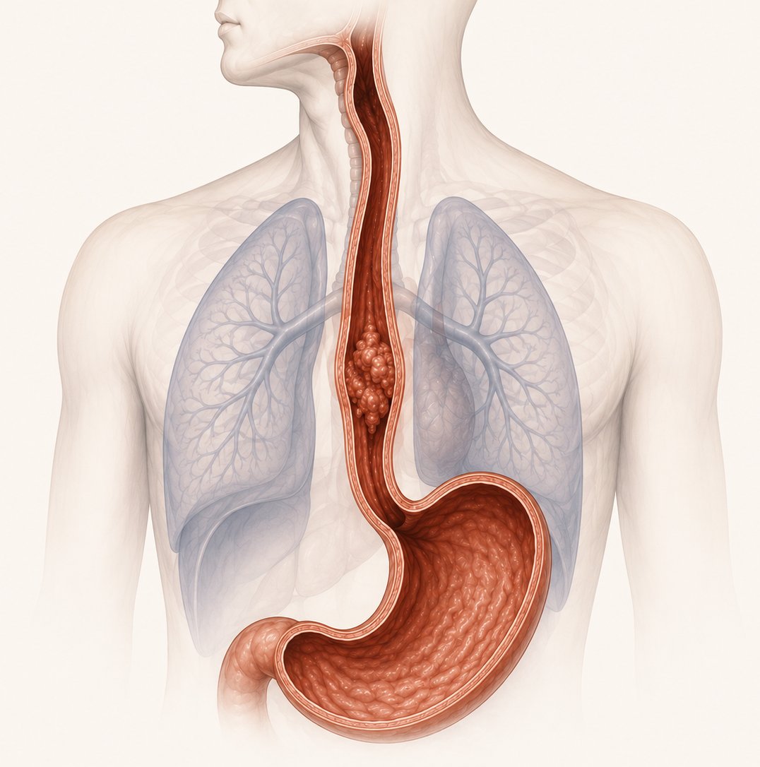
A cancer that begins in the lining of the esophagus — the tube connecting mouth to stomach. It appears as one of two types, each with its own cause, its own location, and its own warning signs.
Two cancers wearing one name
Both begin in the lining of the esophagus, but they arise from different cells, in different places, for different reasons. Tap either to see how they differ.
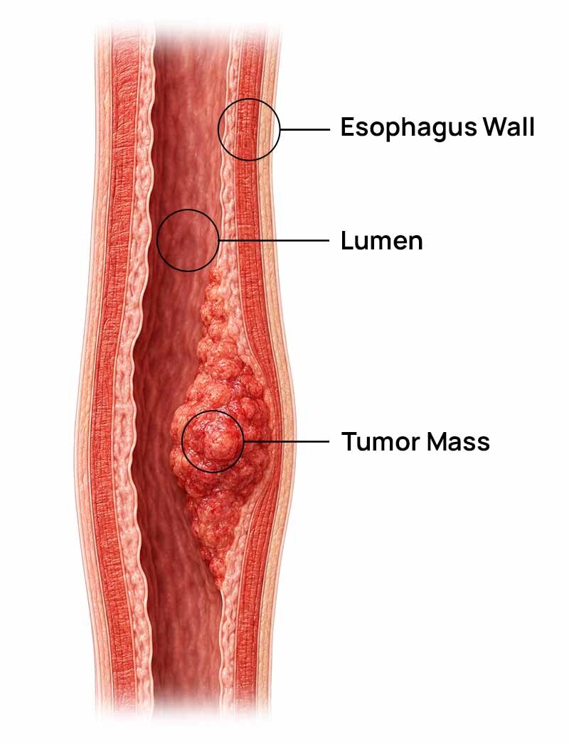
Squamous cell carcinoma
Arises from the flat squamous cells that line the upper two-thirds of the esophagus. Driven by chronic irritation — tobacco, alcohol, very hot drinks and long-term nutritional deficiency. This is the dominant type across India, East Asia and East Africa.
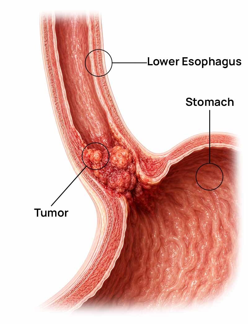
Adenocarcinoma
Arises from glandular cells in the lowest part of the esophagus, where it meets the stomach. Almost always the end point of long-standing acid reflux, which changes the lining into intestinal-type tissue — a condition called Barrett's esophagus. Rising fast wherever obesity and reflux are rising.
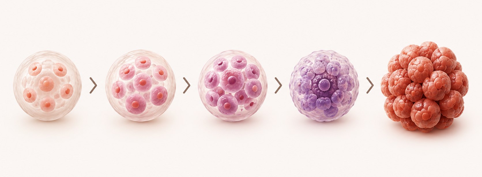
Neither type appears overnight
Both move through a recognisable sequence of tissue changes before becoming invasive. Every step before the last one is detectable — and treatable — which is the entire argument for surveillance.
ESCC pathway
Chronic irritation of the squamous lining — tobacco, alcohol, hot drinks, poor nutrition
Dysplasia — abnormal cell changes appear
Squamous cell carcinoma
EAC pathway
Chronic acid reflux (GERD)
Barrett's esophagus — lining changes to intestinal-type tissue
Low-grade, then high-grade dysplasia
Adenocarcinoma

Who is at higher risk
Most of what raises risk here is either behavioural or treatable. Filter by category.
What to watch for
Tick anything you have noticed. This builds a list you can take to your doctor — it is a note-taking tool, not a test, and it cannot tell you whether you have cancer.
Not sure whether this needs checking?
Talk to our team about what to ask your doctor and which test usually comes first. Free, and there is no obligation to start anything with us.

How it is confirmed
One test proves it. The rest establish how far it has gone.
Upper GI endoscopy with biopsy
A camera passed down the esophagus, with a tissue sample taken from anything suspicious. This is the only test that confirms the diagnosis.
Endoscopic ultrasound
Shows how deeply the tumour has grown into the wall, and whether nearby lymph nodes are involved.
CT chest and abdomen
Assesses local spread and looks for distant metastasis.
PET-CT
More sensitive for distant metastasis and nodal involvement than CT alone.
Bronchoscopy and laparoscopy
Bronchoscopy rules out airway invasion when the tumour sits near the airway. Laparoscopy stages tumours at the gastroesophageal junction.
Staging, plainly
Staging describes how deep the tumour has grown and how far it has travelled. It is the single strongest influence on how care is planned, and it is decided by your medical team — not by a website.
Still only on the surface
Abnormal cells sit in the epithelium, the very top layer of the lining. Nothing has grown deeper and nothing has spread. This stage is usually found by surveillance endoscopy rather than by symptoms.
How it is treated
The approach depends on stage, location and overall health, and usually combines more than one of the below.
Endoscopic therapy
For very early, mucosal disease — endoscopic mucosal resection or radiofrequency ablation.
Esophagectomy
Removal of the affected segment with lymph node dissection, for localised disease.
Chemotherapy
Platinum-based regimens, given before or after surgery, or for advanced disease.
Radiation
Often combined with chemotherapy for locally advanced or inoperable tumours.
Targeted & immune therapy
Anti-HER2 agents for HER2-positive disease; checkpoint inhibitors for PD-L1-positive advanced disease.
Symptom management
Esophageal stenting, feeding tube support and palliative radiation.
Ayurveda's place is alongside, never instead
Used traditionally to support strength, digestion and quality of life during treatment and recovery. It does not treat the cancer, and it does not replace anything your oncology team has planned.
Traditional restorative practices used to support general strength and recovery alongside medical treatment.
Food guidance for a narrowed esophagus and a treatment-strained gut — softer textures, smaller meals, adequate calories.
Routine, rest and stress-management practices to support overall wellbeing through a long treatment course.
Guidance offered in coordination with your oncology team, with a written summary you can hand to them.
A conversation, not a commitment
Three quick questions, then we call you back. Free, confidential, and never a request to stop your treatment.
Thank you — we have your request
One of our team will call you back within one working day. If it is urgent, please call us directly.
What actually lowers risk
- ✓Avoid tobacco in all forms and limit alcohol
- ✓Manage reflux; get surveillance if Barrett's is present
- ✓Maintain a healthy body weight
- ✓Eat more fruit and vegetables
- ✓Let very hot food and drinks cool first
- ✓Treat achalasia and other underlying conditions
- ✓Reduce processed, spicy and pickled foods
- ✓Smaller, more frequent meals; don't lie down after eating
Answered honestly
The two main types are squamous cell carcinoma, which arises in the upper and middle esophagus, and adenocarcinoma, which arises in the lower esophagus and is usually linked to long-standing reflux and Barrett's esophagus.
The most common presenting symptom is progressive difficulty swallowing — first with solids, later with liquids. Because it usually appears only once the tumour has narrowed the esophagus, it is a late sign rather than an early one, and should be evaluated promptly rather than monitored.
Upper GI endoscopy with biopsy is the gold standard, confirming the diagnosis through direct tissue sampling. Endoscopic ultrasound, CT of the chest and abdomen, and PET-CT then establish how far the disease has spread.
As with any major surgery, risks include infection, bleeding and complications with healing at the surgical site. Esophagectomy is a significant operation, and your surgical team will discuss individual risk based on your health and stage.
No. Ayurveda is not a cure for esophageal cancer and should never replace oncology treatment. It is used only as supportive care alongside medical treatment — to help with strength, digestion and general wellbeing — and should be coordinated with your treating oncologist.
Related guides
References
- National Cancer Institute — Esophageal Cancer. cancer.gov
- American Cancer Society — Esophagus Cancer. cancer.org
- World Health Organization — Cancer fact sheet. who.int
- IARC Global Cancer Observatory. gco.iarc.fr
This page is for general information only and does not replace diagnosis or advice from a qualified oncologist. Ayurvedic supportive care described here is a complement to, not a substitute for, medical treatment. Always consult your treating physician before making changes to your care plan.
Your first step is a conversation, not a commitment.
Bring your reports. Talk to our team. We will tell you honestly what supportive care could and could not do for your situation — before you decide anything.